Animal Cheek Cell Under Microscope : Mouth Cells / Smear the cotton swab on a slide (you won't see much without the microscope, but it is probably there.) and place a slide cover over that area.
byLeonard Bodrey-
0
Animal Cheek Cell Under Microscope : Mouth Cells / Smear the cotton swab on a slide (you won't see much without the microscope, but it is probably there.) and place a slide cover over that area.. Epithelial cheek cells ks3 cell plant cell. Label the following parts of human cheek cell brainly in. You can see what it might look like in the image above. Comparing animal and plant cell. To look at a cell close up we need a microscope.
These are plant cells of a leaf of elodea. Students will draw what was visualized to record their observations. The cotton bud should then be smeared onto the slide, leaving residue on the slide. Methylene blue solution can stain the cells with blue color so they are easier to see under the microscope. Which of the following has prokaryotic cells?
Chris Sacchi And Kurt Friehauf Animal And Plant Cells With Elementary School Students from faculty.kutztown.edu Cheek cells under a microscope requirements, preparation and staining cheek cells are eukaryotic cells (cells that contain a nucleus and other organelles within enclosed in a membrane) that are easily shed from the mouth lining. As such it is a favorite in biology classrooms to show what a typical animal cell looks like. Find out how to observe cells under a microscope Neha observed a slide of human cheek cells under a microscope in its i low magnifying power ii high magnifying power settings. This eliminates which type of cell? Which of the following has prokaryotic cells? Use cleandry mounted slide while placing it under the lens of the microscope. An organism with a nucleus c.
Mitochondrion) are visible with a light microscope but can't be seen in detail.
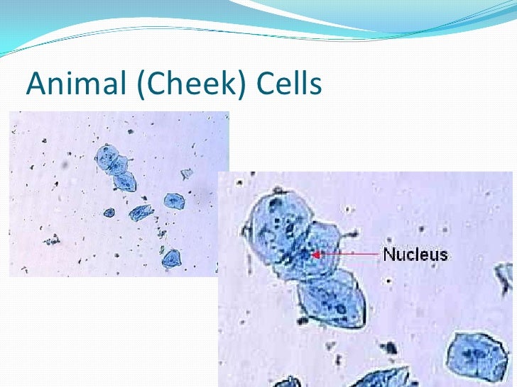
Animal cells tend to lack cell walls and chloroplasts, while plant cells do contain chloroplasts and have cellulose cell walls. Rub a cotton swab across your cheek. This dye is toxic when ingested and it causes irritation when in contact with the skin and eyes. The dye will allow you to clearly stain the nuclei of the cells. You may ask, why is chloroplast not visible in onion cells? Can you id this long stringy material seen in an animal cheek cell. (you should observe the cell membrane, nucleus, and cytoplasm.) Note the nucleus and the numerous mitochondria. These cells line the buccal cavity in humans and are usually shed during mastication and even talking. Methylene blue solution can stain the cells with blue color so they are easier to see under the microscope. Human cheek cells figure 3. Cheek cells 400x these are cheek cells stained with meth flickr. The cheek cell, an example of an animal cell, generally has a circular, oval shape.
Step 3 cover the sample with a drop of water. Can you id this long stringy material seen in an animal cheek cell. The cheek cell, an example of an animal cell, generally has a circular, oval shape. Peptidoglycan cell walls are found. An organism with a nucleus c.
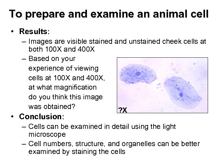
Cell Microscope Lab from image.slidesharecdn.com The onion epidermis cell is the only cell that has a cell wall. Process for obtaining, staining, and viewing cheek cells using a microscope Structure and function of cells learn biology class 8. Click to see full answer. Epithelial cheek cells observed with tomographic diffractive. Also note the ovoid irregular shape of the cell reflecting an absence of a cell wall. Smear the cotton swab on the center of the slide for 2 to 3 seconds. 4.2 (a) to prepare and examine one animal cell, stained and unstained, using the light microscope step 1 swab the inside of the cheek.
Methylene blue solution can stain the cells with blue color so they are easier to see under the microscope.
Label the following parts of human cheek cell brainly in. Human cheek cell under microscope diagram. Draw a diagram of one cheek cell and label the parts. Smear the cotton swab on the center of the slide for 2 to 3 seconds. An organism with a nucleus c. Cheek cells are easy to obtain and easy to see under a microscope. They will be used today for you to observe a eukaryotic animal cells and its nucleus. Step 3 cover the sample with a drop of water. Cheek cells under a microscope requirements, preparation and staining cheek cells are eukaryotic cells (cells that contain a nucleus and other organelles within enclosed in a membrane) that are easily shed from the mouth lining. Animal cells this lesson looks at the structure of an animal cell, how to prepare a human cheek cell slide to view under a microscope and calculating magnification. Due to the fact that the cheek cell was not in groups or clumps, the arrangement of this type of cell is unknown. The cells in the cheeks are eukaryotic cells with a defined nucleus enclosed inside a nuclear membrane along with other cell organelles. Cheek cells are constantly shed from the buccal mucosa, and divide every 24 hours to compensate for this.
Human cheek cells figure 3. Under a microscope, a cell appears to have a nucleus. Add a drop of iodine solution and add a coverslip. This dye is toxic when ingested and it causes irritation when in contact with the skin and eyes. The dye will allow you to clearly stain the nuclei of the cells.
Chapter 6 Cell Structure Leaving Certificate Biology Higher from present5.com Peptidoglycan cell walls are found. Cheek cells under a microscope requirements, preparation and staining cheek cells are eukaryotic cells (cells that contain a nucleus and other organelles within enclosed in a membrane) that are easily shed from the mouth lining. To create the cheek cells slide, use the flat end of a toothpick to gently scrape the inside of your cheek, then spread the cells on a clean slide. The cells in the cheeks are eukaryotic cells with a defined nucleus enclosed inside a nuclear membrane along with other cell organelles. Human cheek cell under microscope diagram. Which of the following has prokaryotic cells? List 3 organelles that were not visible but should have been in the cheek cell. You will scrape and stain a sample of your cheek cells with the dye methylene blue.
Click to see full answer.
Structure and function of cells learn biology class 8. It's therefore easy to obtain them for observation. Shop' die neuesten kollektionen bequem online bei surfdome jetzt. Use unusednew toothpick for scraping of cheek cells. The cotton bud should then be smeared onto the slide, leaving residue on the slide. Smear the cotton swab on a slide (you won't see much without the microscope, but it is probably there.) and place a slide cover over that area. Add a drop of methylene blue solution on the smear. Microscope students need to gently wipe the lining of the inside of their own cheek with the end of a sterile cotton bud. 4.2 (a) to prepare and examine one animal cell, stained and unstained, using the light microscope step 1 swab the inside of the cheek. Step 3 cover the sample with a drop of water. You can see what it might look like in the image above. Find out how to observe cells under a microscope This will gather some cells from the lining of the cheek wall.