Animal Cheek Cell Under Microscope / 1 Lab Plant And Animal Cells - (you should observe the cell membrane, nucleus, and cytoplasm.)
byLeonard Bodrey-
0
Animal Cheek Cell Under Microscope / 1 Lab Plant And Animal Cells - (you should observe the cell membrane, nucleus, and cytoplasm.). Then view at higher magnification. 4.2 (a) to prepare and examine one animal cell, stained and unstained, using the light microscope step 1 swab the inside of the cheek. Human cheek cells are made of simple squamous epithelial cells, which are flat cells with a round visible nucleus that cover the inside lining of the cheek.c. Specifically epithelial cells from the inside of. Under a microscope, plant cells from the same source will have a uniform size and shape.
Instructions for collecting cheek cells and mounting on slide: The vacuole in an an animal cell is smaller. Cheek cells are constantly shed from the buccal mucosa, and divide every 24 hours to compensate for this. Step 3 cover the sample with a drop of water. Human cheek cells under microscope human cheek cells 400x.
Observation Of Methylene Blue Stained Cheek Cells Using Foldscope Microcosmos from i2.wp.com Methylene blue stains negatively charged molecules in the cell, including dna and rna. Cell wall, nucleus and chloroplasts can be seen with a compound light microscope under a total magnification of 400 x. Use cleandry mounted slide while placing it under the lens of the microscope. Then view at higher magnification. Epithelial cheek cells observed with tomographic diffractive. This cell do not have plastids, vacuoles or cell wall. As such it is a favorite in biology classrooms to show what a typical animal cell looks like. An rna stain like pyronin y might highlight the nucleolus if there is much of one to see, but i've never tried that.
To obtain the cheek cells you'll use for observation under the microscope, you'll need a toothpick.
Cheek cells are easy to obtain and easy to see under a microscope. They have irregular cellular thin boundaries which contains jelly like cytoplasm and the cytoplasm are granular. Observing plant and animal cells. Specifically epithelial cells from the inside of. Human cheek cell under microscope diagram. Instructions for collecting cheek cells and mounting on slide: You can see what it might look like in the image above. Neha observed a slide of human cheek cells under a microscope in its i low magnifying power ii high magnifying power settings. Smear the cotton swab on a slide (you won't see much without the microscope, but it is probably there.) and place a slide cover over that area. In addition, it is the only cell that has a chloroplast, where photosynthesis can happen. This cell do not have plastids, vacuoles or cell wall. 1) add one drop of food coloring to the middle of a clean slide. Step 2 transfer the sample to a slide.
It's therefore easy to obtain them for observation. 4.2 (a) to prepare and examine one animal cell, stained and unstained, using the light microscope step 1 swab the inside of the cheek. The onion epidermis cell is the only cell that has a cell wall. Have the students collect their own cheek cells to prepare slides for viewing under the microscope. Within the cell, there is a shape of round with a circular structure of granulated part on the epithelial cells.
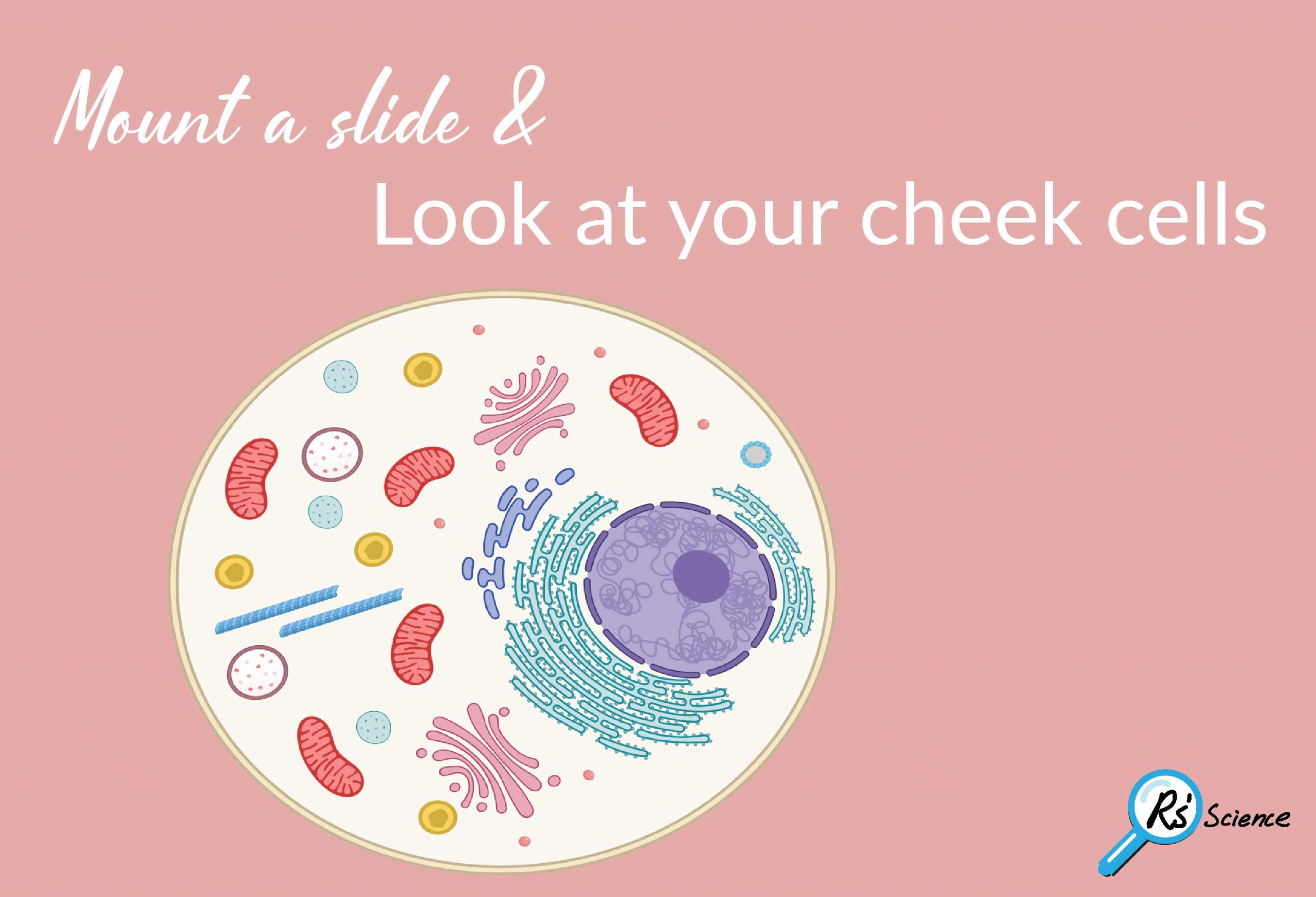
Lesson 2 Mount A Slide Look At Your Cheek Cells Rs Science from rsscience.com Under a microscope, plant cells from the same source will have a uniform size and shape. In addition, it is the only cell that has a chloroplast, where photosynthesis can happen. Produkte für gewerbe und wissenschaft. Human cheek cell under microscope diagram. Epithelial cells surround the internal surface of the mouth which can be taken out using finger nails or a small spoon. Human cheek cells under the microscope w/ commentary. (you should observe the cell membrane, nucleus, and cytoplasm.) Also, like the cheek cell, the onion skin cells were pushed together so that no spaces were in between.
Neha observed a slide of human cheek cells under a microscope in its i low magnifying power ii high magnifying power settings.
Due to the fact that the cheek cell was not in groups or clumps, the arrangement of this type of cell is unknown. You can observe this epithelial animal cell under microscope with high power. Step 3 cover the sample with a drop of water. 1) add one drop of food coloring to the middle of a clean slide. Specifically epithelial cells from the inside of. Beneath a plant cell's cell wall is a cell membrane. Epithelial cheek cells observed with tomographic diffractive. Process for obtaining, staining, and viewing cheek cells using a microscope It's therefore easy to obtain them for observation. Human cheek cell under microscope diagram. Rub a cotton swab across your cheek. Human cheek cells are made of simple squamous epithelial cells, which are flat cells with a round visible nucleus that cover the inside lining of the cheek. Cheek cells under a microscope requirements, preparation and staining cheek cells are eukaryotic cells (cells that contain a nucleus and other organelles within enclosed in a membrane) that are easily shed from the mouth lining.
Under a microscope, plant cells from the same source will have a uniform size and shape. Specifically epithelial cells from the inside of. To obtain the cheek cells you'll use for observation under the microscope, you'll need a toothpick. As such it is a favorite in biology classrooms to show what a typical animal cell looks like. Draw a diagram of one cheek cell and label the parts.
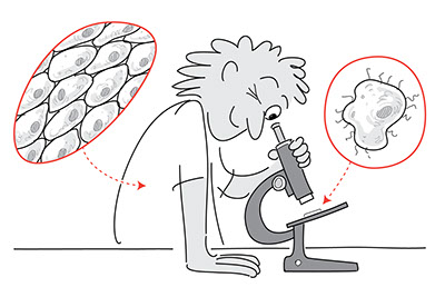
Lab Using A Microscope from serpmedia.org Also, like the cheek cell, the onion skin cells were pushed together so that no spaces were in between. As such it is a favorite in biology classrooms to show what a typical animal cell looks like. We zoom in on an individual cell at 28:00we look at the cheek cells through 40x, 100x, 400x, and x1000. Cell wall, nucleus and chloroplasts can be seen with a compound light microscope under a total magnification of 400 x. Specifically epithelial cells from the inside of. Epithelial cells surround the internal surface of the mouth which can be taken out using finger nails or a small spoon. Epithelial cheek cells observed with tomographic diffractive. Cheek cells under a microscope requirements, preparation and staining cheek cells are eukaryotic cells (cells that contain a nucleus and other organelles within enclosed in a membrane) that are easily shed from the mouth lining.
Within the cell, there is a shape of round with a circular structure of granulated part on the epithelial cells.
Epithelial cheek cells observed with tomographic diffractive. Step 3 cover the sample with a drop of water. Neha observed a slide of human cheek cells under a microscope in its i low magnifying power ii high magnifying power settings. How to make a slide to view cheek cells under the microscope. Cheek cells are easy to obtain and easy to see under a microscope. Step 2 transfer the sample to a slide. Place your slide under the microscope and make the adjustments to see the individual cheek cells. Cheek cells under a microscope requirements, preparation and staining cheek cells are eukaryotic cells (cells that contain a nucleus and other organelles within enclosed in a membrane) that are easily shed from the mouth lining. Under a microscope, plant cells from the same source will have a uniform size and shape. This dye is toxic when ingested and it causes irritation when in contact with the skin and eyes. Within the cell, there is a shape of round with a circular structure of granulated part on the epithelial cells. Also, like the cheek cell, the onion skin cells were pushed together so that no spaces were in between. They have irregular cellular thin boundaries which contains jelly like cytoplasm and the cytoplasm are granular.
How to make a slide to view cheek cells under the microscope cheek animal cell microscope. Due to the fact that the cheek cell was not in groups or clumps, the arrangement of this type of cell is unknown.